Wednesday, May 7, 2014

what is the cause for this condition and your plan of management?

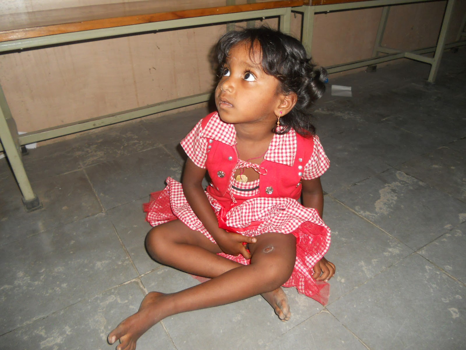
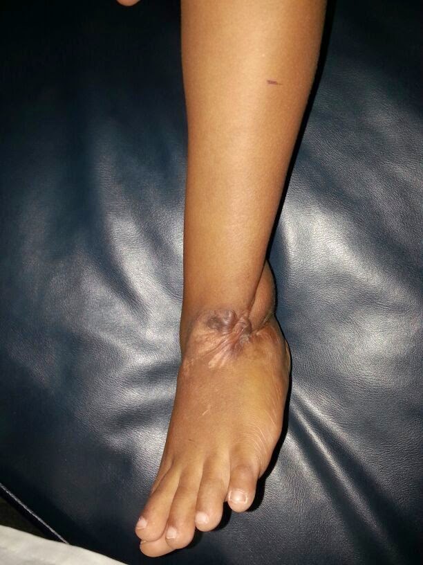
a 14 yr old boy came with chief complaints of deformity of rt forearm
h/o trauma 6 years ago..was hit by teacher at school
later he was taken to an outside hospital where the reports suggest he had been operated for compartment syndrome and the operative notes say that they found pockets of pus

o/e
valgus deformity at forearm
midline healed surgical incision
movements are normal except ulnar deviation at wrist

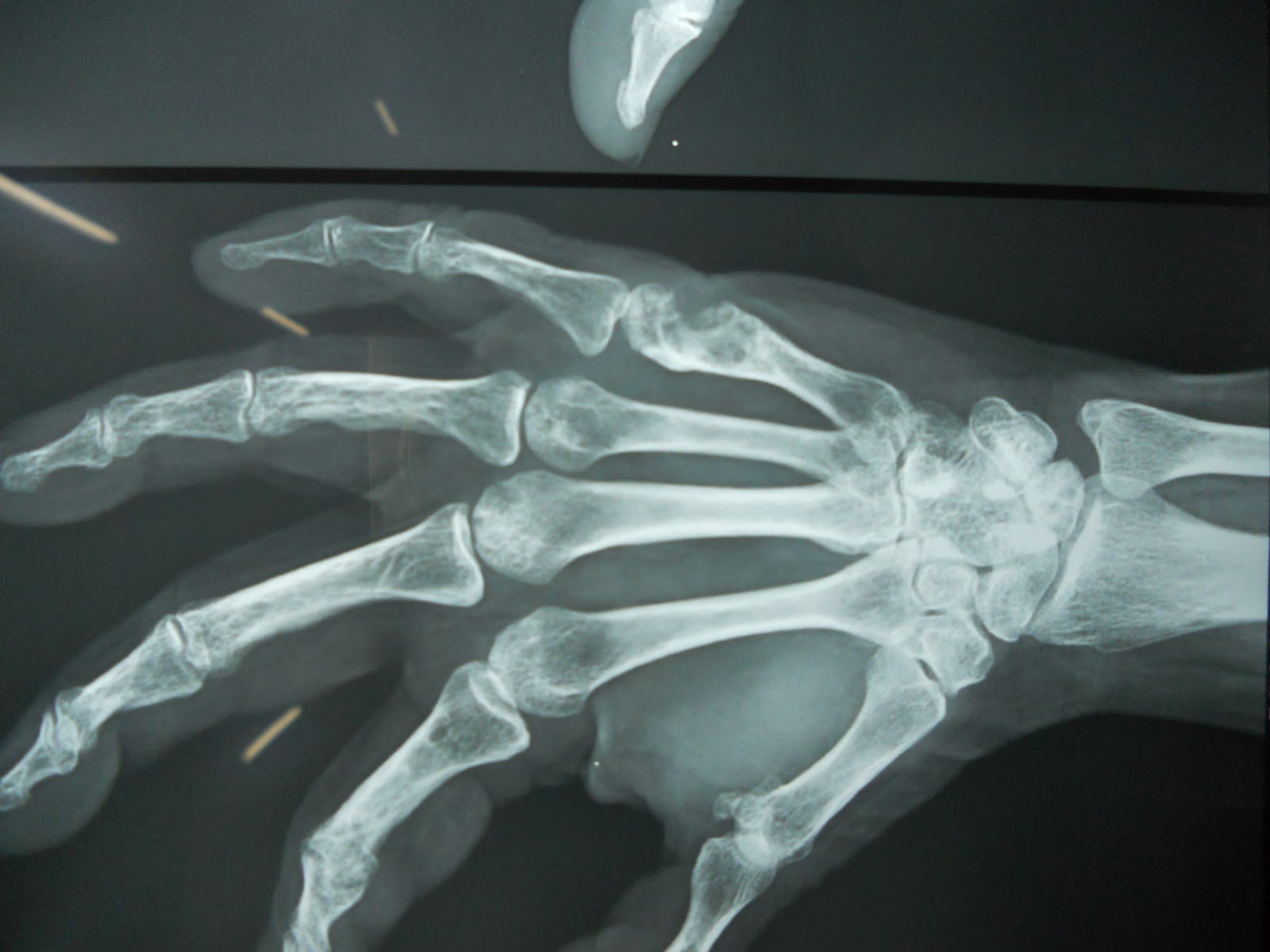
x-ray

Saturday, March 8, 2014

clinical case

a 30 year old male came with chief complaints of pain in hip since 1 year.swelling since 6 months
no relevant trauma
no weight loss
no loss of appetite
O/E: 40 degree of fixed flexion deformity
         swelling seen and palpable anteriorly and posteriorly
          ROM very painful
what is ur radiological diagnosis
what is ur approach to this case

Friday, October 5, 2012

case 8


A 20 year old female patient presented to our OPD with 
complaints of pain in her left foot since about 10 months and 
swelling in her left foot since 3 months. 


HOPI 

Patient was apparently asymptomatic 10 months back when she developed pain in her foot which is insiduos in onset, of dull aching in nature, aggravated by standing and walking, and relieved by taking rest and/or medication. Pain is of increasing severity and is continuous  since last 2 months. 

Patient developed swelling in her left foot about 3 months back initially diffuse and gradually localized to mid dorsum of foot. There is no history of increase in the size of the swelling.

No h/o trauma
No h/o fever/ weight loss / constitutional symtoms.
No h/o numbness or paraesthesias in the foot.
Past History

No h/o similar complaints i the past.
No h/o TB/ DM/ HTN/ Asthma/ epilepsy/ bleeding disorders.
No h/o past hospitalisation/ surgery


Personal History

Diet mixed
Appetite normal
Sleep disturbed of late due to pain


Family History:

No h/o similar complaints in family members.
No h/o any tuberculous contact.


General examination
20 yr old female who is c/c/c, moderately built and nourished.with pallor,and is without icterus, cyanosis, clubbing,  generalized lymphadenopathy, and pedal edema.
Gait antalgic
Vitals stable
other systems normal

Local examination of her left foot
Inspection


A globular swelling of 3cms diameter is noted over the dorsum of her left mid foot.


swelling is diminishing in size on extension of toes and surface is smooth ,skin over the swelling is normal, no scars and sinuses no visible pulsations, and no local inflammatory signs noted. surroundings are normal.


Palpation:
Local warmth noted,
Deep tenderness noted over the cuneiform bones and the bases over first second third metatarsal.
Swelling site and size are confirmed,skin over the swelling is pinch able and the swelling is uniformly soft  in consistency fluctuant , margins well defined and has minimal mobility in AP and lateral directions. No translucency, neither reducible nor compressible.


Workup and radio graphs attached
MR will be attached shortly


9 months old x rays


Present X rays


Histopathology report of Open biopsy attached


Aspiration yielded straw colored thin fluid which was sterile on microscopy and culture
Open biopsy yielded bits of cheesy material and bony spicules of medial and middle cunieform bones.

What is your clinical radio logical and final diagnosis
how would you like to further manage this patient?


Wednesday, May 23, 2012

clinical case 7


A 3 and half year old child came for review in opd after 2 years for follow up and review...

Child at the age of 1and1/2 yrs developed DVT of her left lower limb following ? DPT vaccination in thigh for which she received parenteral heparin. Following which hematoma developed in left thigh which got infected in due course of illness got converted to abscess with discharging sinus. ! year following the persistent discharging sinus a x ray  of left thigh showed features suggestive of COM left femur with sequestrum. The child was treated in this hospital for COM with sequestrectomy and saucerisation. 

Now the present x rays and clinical photographs show the remarkable recovery of the child and x ray with hardly any evidence of previous pathology.

Wednesday, May 16, 2012

Friday, March 2, 2012

Case 6

 A 9yr old male child was presented by his mother in our
      opd with complaints of

      pain in the right knee since two years and

      difficulty in walking since two years

    HOPI:      (Informant is mother)
    
        Pt was apparently alright 2 years back when the
      child injured his right knee while playing for which no
      medical advice was sought following which child developed
      limp and pain while walking
    
      Pain is localized to right knee which is dragging type which
      is aggravated by activity and relieved by rest. No history of
      radiation.
    
      Mother noticed limp while her child walks and has difficulty
      walking and running after the event which is non-progressive.
    
      No h/o similar complaints in the past.
    
      no h/o similar complaints in family members.
    
      Patient was operated at a local government hospital 4months
      back for his condition with no improvement

    
On General Examination:    
      A 9 yr old male child who is moderately built and moderately
      nourished who is afebrile and is otherwise healthy with
      stable vitals 

 Local examination:

    Decubitus:
      Pt is lying comfortably on couch with umbilicus central both
      ASISs at same level with both hips in neutral position with
      apparent wasting in thigh component with right patella facing
      the wall left facing the roof both knees in neutral position
      and foot plantigrade.
    Inspection:
    
      On inspection a surgical healed scar of 5cms long is noted
      over the lateral aspect of right knee and a post traumatic
      healed scar of 1X1 cm noted in the supero-medial aspect of
      right knee. The medial aspect of the knee is more prominent
      in comparision to opposite knee. No swellings are noted. No
      Parapatellar fossa fullness is noted. Patella appears to be
      laterally displaced in comparision to opposite knee. No
      sinuses noted.

   
Palpation:
    
      No local rise of temperature
      tenderness is present in the joint line of the right knee and
      also elicited in the patellofemoral joint and lateral margin
      of patella. Patella is found to be subluxated and its more
      mobile laterally compared to opposite knee. The medial
      prominence is continuous with the femoral medial condyle
      whose size is comparable to the opposte knee. Inspectory
      findings of scars and parapatellar fossae are confirmed.

    Movements:
    
      patient is having a fixed flexion deformity of 10 degrees
      with further full range of flexion active and passive
      possible with terminal 10 degrees of flexion painful.
    
      Patient is flexing abducting and external rotating his right
      hip to extend the knee and dragging the foot over the crouch
      to actively extend his knee.
    
      The patella is dislocating laterally with every flexion of
      right knee.

   
Measurements
      
          The size of the patella is comparable to opposite side. There
      is two inches wasting noted in right thigh component.

   
Gait:
    
      child avoids flexion of knee while walking and running

SPECIAL  TESTS    Q Angle
      measured in 30deg knee flexion is 20 degrees

    Apprehension test is positive as the patient avoids flexion of kneewhen lateral
      dislocating pressure is applied over the patella.

    Patellar tilt test is positive

Radiological examination:
    
      x rays of right knee AP & Lat suggestive of normal sized
      patella with hypoplastic lateral condyle of rt femur. Patella
      on axial projections shows flattened lateral n medial
      articulate facets with decrease in size of lateral articular
      facet. Standing x rays suggestive of horizontal joint line
      and the decrease in size of lateral femoral condyle is
      confirmed.
    
      Insall ratio is around 1.5

    Diagnosis:
    
      Recurrent dislocation of Right patella in 9 years old pt.


      Kindly reviews the clinical pictures, videos and radiographs 
       and suggest the optimal management for the child's
      condition.


AP view

Lat view shows lat condyle hypoplasia


Axial view of patella shows small lateral articular facet


X ray both knees AP in standing


Displaced patella in flexion


Lateral scar