Wednesday, May 23, 2012

clinical case 7


A 3 and half year old child came for review in opd after 2 years for follow up and review...

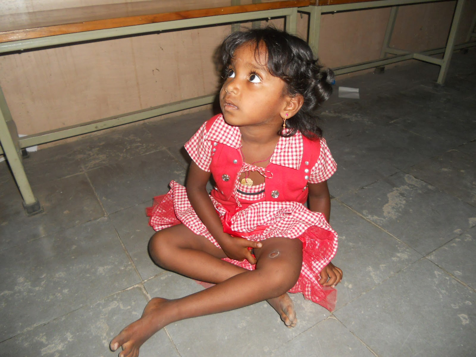
Child at the age of 1and1/2 yrs developed DVT of her left lower limb following ? DPT vaccination in thigh for which she received parenteral heparin. Following which hematoma developed in left thigh which got infected in due course of illness got converted to abscess with discharging sinus. ! year following the persistent discharging sinus a x ray  of left thigh showed features suggestive of COM left femur with sequestrum. The child was treated in this hospital for COM with sequestrectomy and saucerisation. 

Now the present x rays and clinical photographs show the remarkable recovery of the child and x ray with hardly any evidence of previous pathology.

Wednesday, May 16, 2012

Friday, March 2, 2012

Case 6

 A 9yr old male child was presented by his mother in our
      opd with complaints of

      pain in the right knee since two years and

      difficulty in walking since two years

    HOPI:      (Informant is mother)
    
        Pt was apparently alright 2 years back when the
      child injured his right knee while playing for which no
      medical advice was sought following which child developed
      limp and pain while walking
    
      Pain is localized to right knee which is dragging type which
      is aggravated by activity and relieved by rest. No history of
      radiation.
    
      Mother noticed limp while her child walks and has difficulty
      walking and running after the event which is non-progressive.
    
      No h/o similar complaints in the past.
    
      no h/o similar complaints in family members.
    
      Patient was operated at a local government hospital 4months
      back for his condition with no improvement

    
On General Examination:    
      A 9 yr old male child who is moderately built and moderately
      nourished who is afebrile and is otherwise healthy with
      stable vitals 

 Local examination:

    Decubitus:
      Pt is lying comfortably on couch with umbilicus central both
      ASISs at same level with both hips in neutral position with
      apparent wasting in thigh component with right patella facing
      the wall left facing the roof both knees in neutral position
      and foot plantigrade.
    Inspection:
    
      On inspection a surgical healed scar of 5cms long is noted
      over the lateral aspect of right knee and a post traumatic
      healed scar of 1X1 cm noted in the supero-medial aspect of
      right knee. The medial aspect of the knee is more prominent
      in comparision to opposite knee. No swellings are noted. No
      Parapatellar fossa fullness is noted. Patella appears to be
      laterally displaced in comparision to opposite knee. No
      sinuses noted.

   
Palpation:
    
      No local rise of temperature
      tenderness is present in the joint line of the right knee and
      also elicited in the patellofemoral joint and lateral margin
      of patella. Patella is found to be subluxated and its more
      mobile laterally compared to opposite knee. The medial
      prominence is continuous with the femoral medial condyle
      whose size is comparable to the opposte knee. Inspectory
      findings of scars and parapatellar fossae are confirmed.

    Movements:
    
      patient is having a fixed flexion deformity of 10 degrees
      with further full range of flexion active and passive
      possible with terminal 10 degrees of flexion painful.
    
      Patient is flexing abducting and external rotating his right
      hip to extend the knee and dragging the foot over the crouch
      to actively extend his knee.
    
      The patella is dislocating laterally with every flexion of
      right knee.

   
Measurements
      
          The size of the patella is comparable to opposite side. There
      is two inches wasting noted in right thigh component.

   
Gait:
    
      child avoids flexion of knee while walking and running

SPECIAL  TESTS    Q Angle
      measured in 30deg knee flexion is 20 degrees

    Apprehension test is positive as the patient avoids flexion of kneewhen lateral
      dislocating pressure is applied over the patella.

    Patellar tilt test is positive

Radiological examination:
    
      x rays of right knee AP & Lat suggestive of normal sized
      patella with hypoplastic lateral condyle of rt femur. Patella
      on axial projections shows flattened lateral n medial
      articulate facets with decrease in size of lateral articular
      facet. Standing x rays suggestive of horizontal joint line
      and the decrease in size of lateral femoral condyle is
      confirmed.
    
      Insall ratio is around 1.5

    Diagnosis:
    
      Recurrent dislocation of Right patella in 9 years old pt.


      Kindly reviews the clinical pictures, videos and radiographs 
       and suggest the optimal management for the child's
      condition.


AP view

Lat view shows lat condyle hypoplasia


Axial view of patella shows small lateral articular facet


X ray both knees AP in standing


Displaced patella in flexion


Lateral scar


Tuesday, January 17, 2012

Case 5


15 years old male patient
Chief complaints:
   pain in the left hip since 8 months.
   limp since 6 months.
History of present illness:
Pateint asymptomatic before,correlate his history to a trivial trauma while playing as he sustained left hip injury due to fall and able to continue game after that,had little discomfort while performing routene activity and started message therapy almost 2 weeks after injury and continuedfor a month.
He developed swelling around the hip and there increase in the intensity of the pain.he was completely bed ridden due to pain. He had high grade fever with chills and rigours.
The swelling increased slowly around the anterior aspect of the left hip and the left iliac region(patient is localizing the site by his finger)patient hospitalized during that course and the surgery done over the left iliac region(scar sugesting of the incision ovr left iliac region)
The pus was drained and further medication given and patient discharged.
The pain subsided after the surgeryto little extent  with limp and then it increased gradually to the present situation,as patient unable to walk to his working place at present
Patient unable to squat and sit crossed leg.
Patient using support while walking or standing from bed since one month.
No history of night fever,weight loss,cough or other contitutional symptoms.
No history of any previous medications.
No history of pain in other joints.
Past history:
history of surgery around 7 months back, as mentioned in present illness.
No history of other illness.
No history of any medications in past.
Personal history:
Diet: mixed
Appetite: normal
Bowel bladder : regular.
Sleep: disturb due to pain.
Addiction: nil.
Family history:
Nothing contributory.
General examination:
Noting contributory.
local examination:
Inspection:
patient lying comfortabily,head central, both shoulder at same level,umblicus central,
Both ASIS at same level, hip in external rotation (L),patella facing laterally(L),foot lateral border touching bed.no exagrated lumbar lordosis.
Scarpa’s triangle free,greater trochanter more prominent than rt side,thigh muscle wasting,
Gluteal muscle wasting,scar over the left iliac fossa(primary intension healing),no sinuses,visible swelling,pulstionsor dilated veins.
No apparent limb length difference.
Palpation:
No local rise of temperature,ASIS at same level,anterior left hip joint line tenderness present,greater trocanter tender,broadened and irregular with no proximal migration.
No palpable mass around hip ant/pos/laterally.
Femoral pulse palpable and symetrical both sides.
Scar over the left iliac fossa is healthy non tender and not adherent to deeper tissue.
Movements:
Left hip:
Flexion: pain free 0 to 60 degrees and further pain full 60 to 100 degrees with axial deviation while flexion.
Extension:0 to 5 degrees of extension pain full
Adduction: 0 to 10 degrees painless and further 10 to 20 degree pain full .
Abduction: 0 to 15 degrees pain less and further 15 to 25 degrees painfull
External rotation : fixed external rotation deformity of 5 degrees with further external rotation of 15 degrees.
Note:pain while terminal range of all movement with palpable crepitus.
Measurments:
Apparent length equall both sides.
True length equal both sides.
Byrants triangle symetrical both sides.
Tests:
Shoe maker line: symetrical meeting at umblicus.
Neletons line: no abnormality.
Chienes parellelogram: normal.
Thomas test: no fixed flexion deformity.
Telescopic test: negative.
Trendelenberg test: positive.
Gait: stable/painfull gait-short limb gait.

Investigations
CBP: WNL
ESR 20mm@ 2ND HR
Montoux Neg
DDX:
septic arthritis of left hip.
Tuberculous  arthritis of left hip.
.
.
.
.


Whats Your DDX?????
How would you proceed further..
Whats your plan of management

Wednesday, November 23, 2011

Case 4

Case of Bilateral AVN

Patient  is a 65 year old male who presented with 
C/o pain in the right hip since 5 years and 
C/o pain in the left hip since 1 month
C/o inability to walk since 1 month

HOPI:
Pt was apparently alright 5 years back when he experienced a fall in his residence, trivial in nature following which he was able to get up and walk though it was painful. Patient took rest at his home and had OTC analgesics. He was alright with in a week  though he experienced pain occasionally on and off with activity. The pain has progressively increased ever since with increasing frequency and severity with disabling intensity since 1 month. Patient experiences pain in the right hip radiating to the right knee. Patient is also experiencing pain in the left hip since 1 month. Patient is unable to sit cross-legged and squat since one month. 
No history of fever
No h/o weight loss
No h/o decreased appetite

O/e: 
Past history:
History of hip surgery in child hood with no any intra/post op complication.
No history of any chronic illness/long term medications.
No history of any allerdies to drugs.
No significant past/personal history and general examination findings.
HIP EXAMINATION:
Inspection:
Patient is examined in supine decubitus
Rightlower limb in 45 degrees flexed position.
Apparently shortened limb.
Right ASIS is at higher level.
Scarpas triangle is free.
Gluteal,thigh muscle wasting present.
no sinuses,swellings,dilated veins or pulsations.
Palpation:
No local rise of temperature,ASIS at higher level at Rt side.
Joint tenderness ant and post.Greater trochanter nontender, relatively lessprominent,poximally migrated,irregular,broadened.
Femur shaft nontender and normal.
Movement:
FFD: 20 DEGREES.
ADDUCTION DEFORMITY:10 DEGREES
EXTERNAL ROTATION:5 DEGREES
INTERNAL ROTATION:5 DEGREES
Measurements:
Apparent shortening 5cms.true shortening 2cms.
2cms wasting of thigh muscles.
2cms supratrochanteric shortening,confirmed by byrants triangle base.
specific tests:
Narath sign: negative.
Nelton’s line trochanter is above the line.
Shoemaker’s linecoinciding on Left of midline below the level of umblicus.

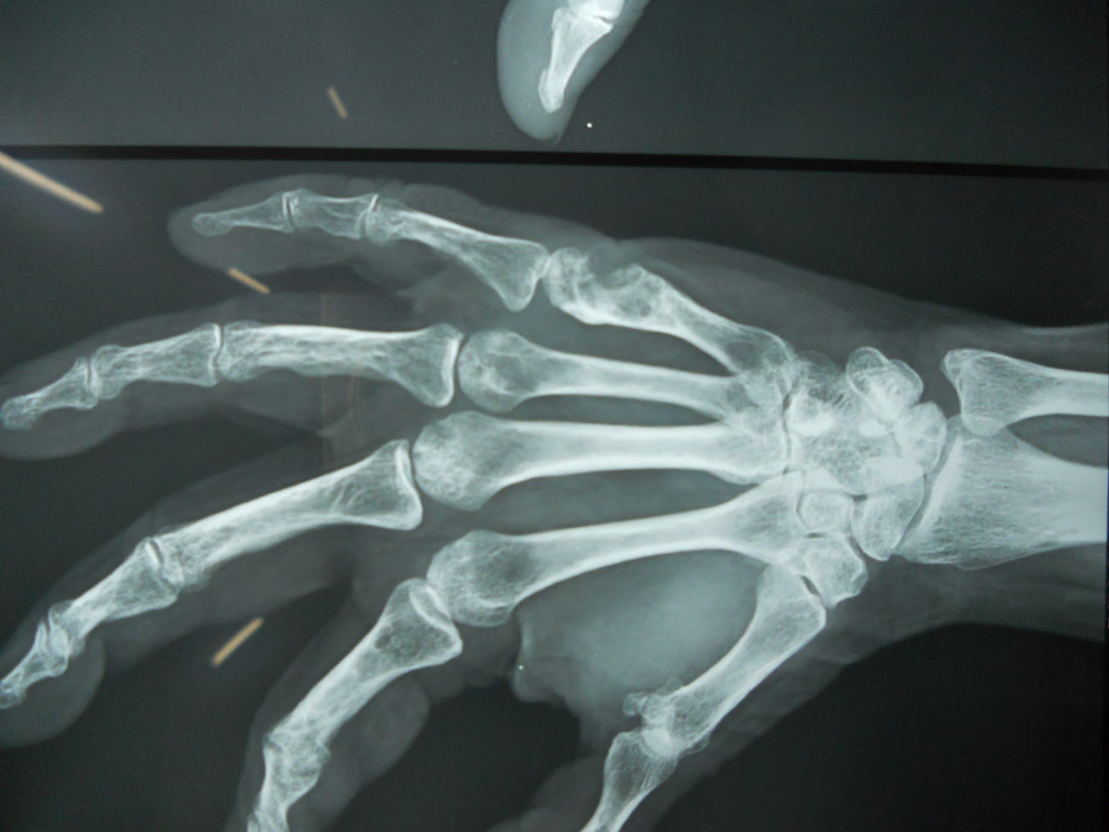
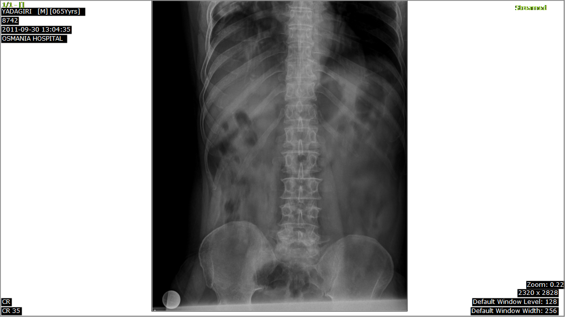
Plain Xray of Pelvis with both hips shows the dense sclerotic and deformed head of Rt femur with changes in acetabulum left hip is apparently normal on X ray

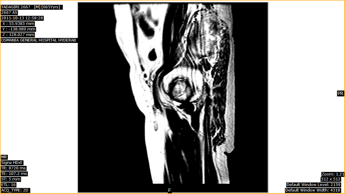
MR Findings


Click on image below to view image n scroll through the series of images in actual size